Ct Elbow Positioning
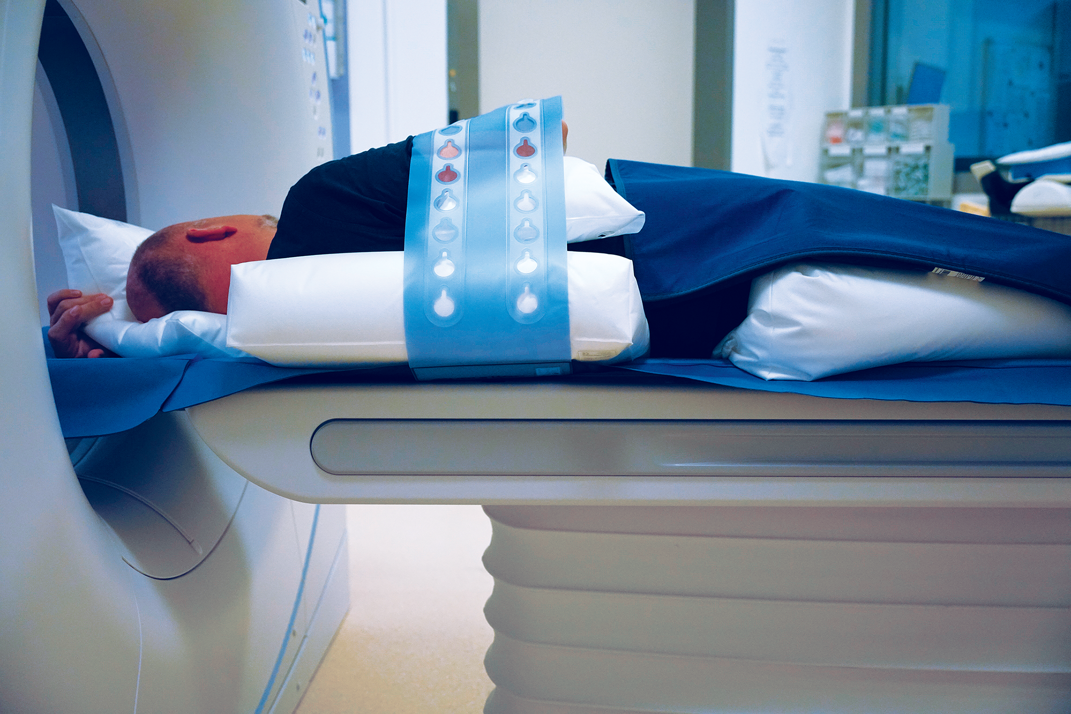
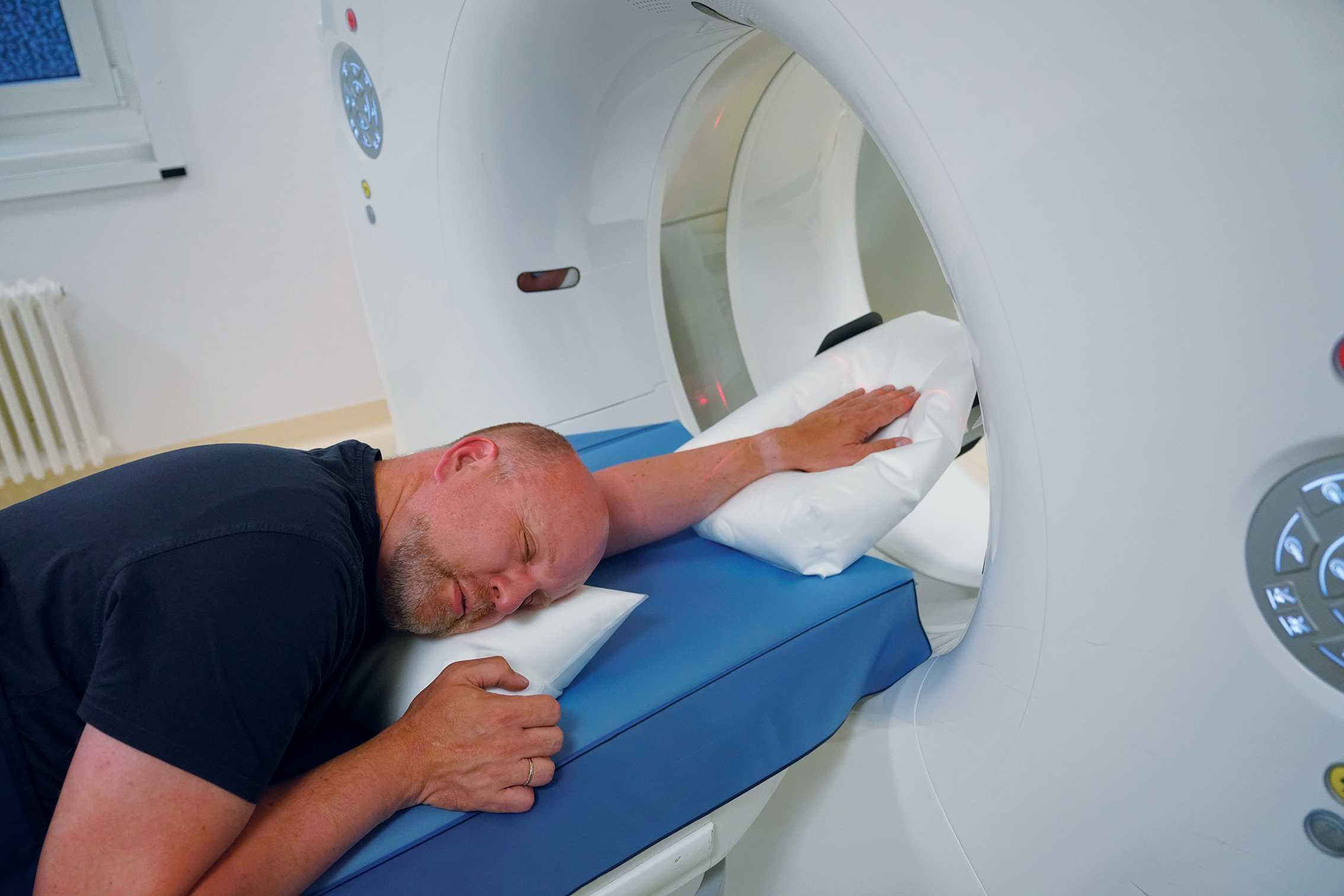
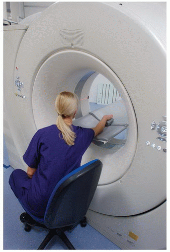
Ct elbow positioning. When assessing a cranio-caudal elbow image the key indicators of good positioning are. - head tucked down and out of scan range. In short-legged dogs it may be necessary to place a pad behind the elbow to push the leg cranially.
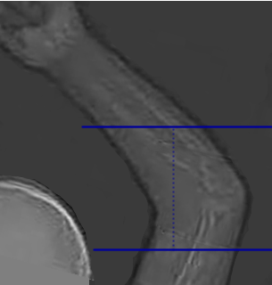
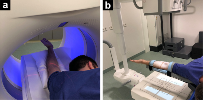
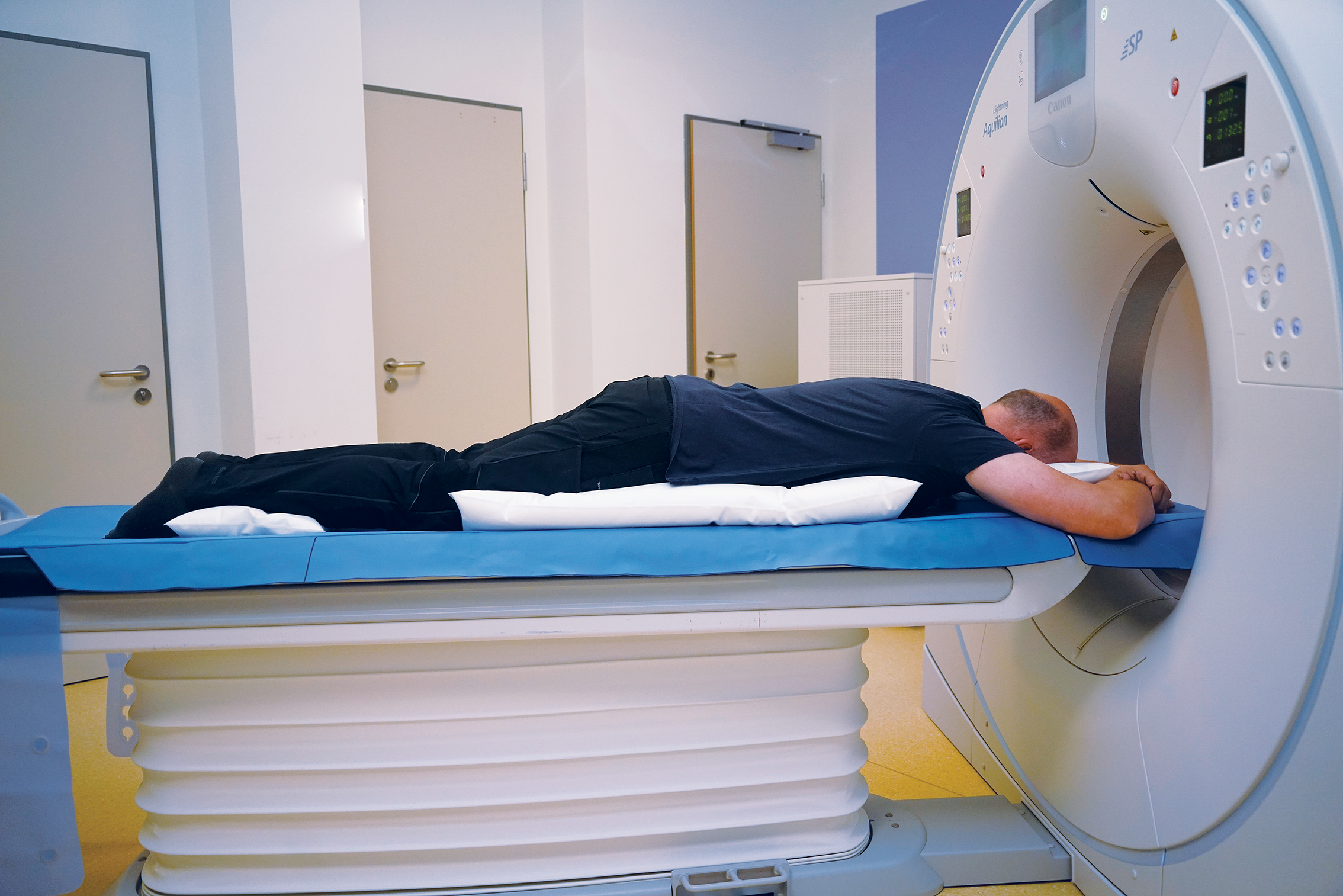
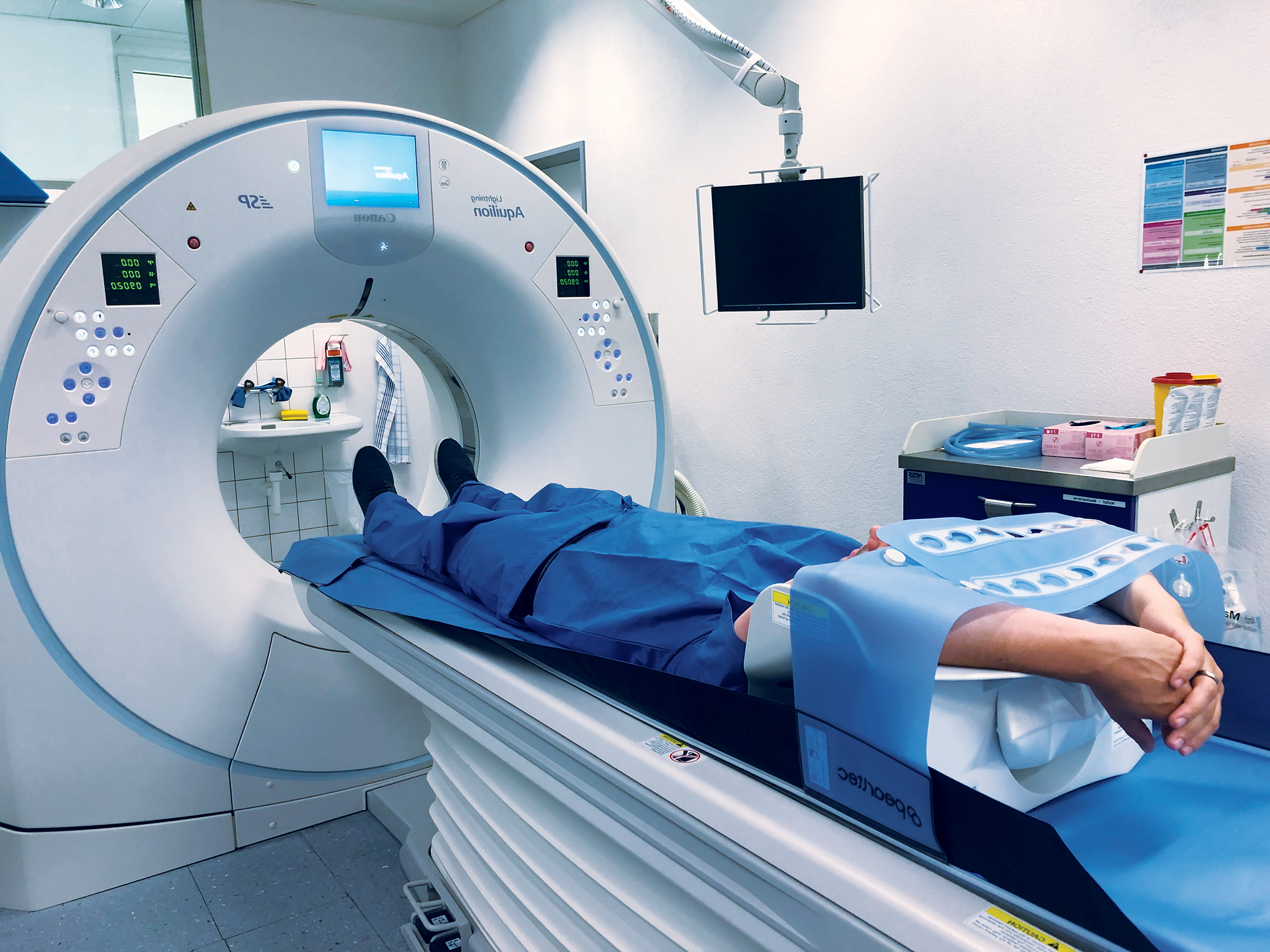
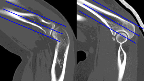
The elbow should be positioned so that the angle between the humerus and radiusulna is 45 avoiding over flexion. - affected arm above head immobilised with sponges and strap. Coronal Imaging Plane Prescribe plane parallel to anterior humerus at condyles.
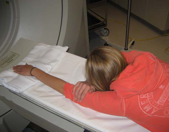
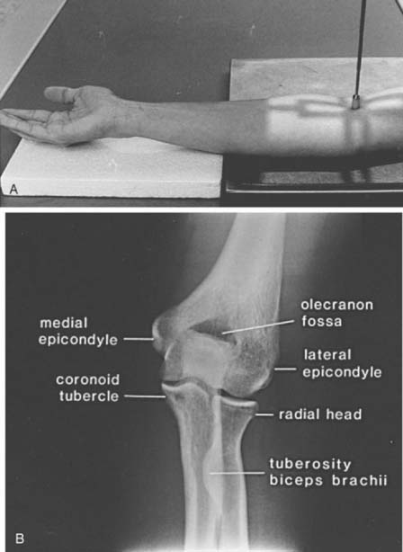
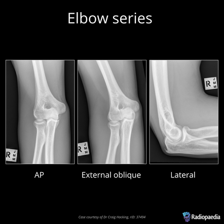
Position of part The arm bent 90 degrees thumb up and the humeral epicondyles should be 90-degrees to the plane of the the image receptor. Place the arm on the table with elbow straight. Can do to optimize Bone CT 1 Optimize Patient Positioning Try to center the bone Get other bonesmetal out of scanning FOV 2 Optimize Scanning Technique.
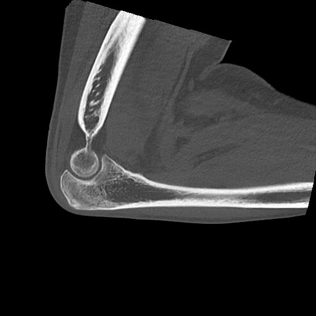
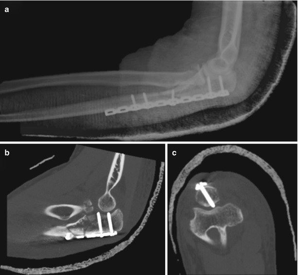
Ct Scan Elbow Positioning. Scan of the elbow after opacification by iodinated contrast from the joint cavity of the elbow. The patient is asked to look away from the gantry.
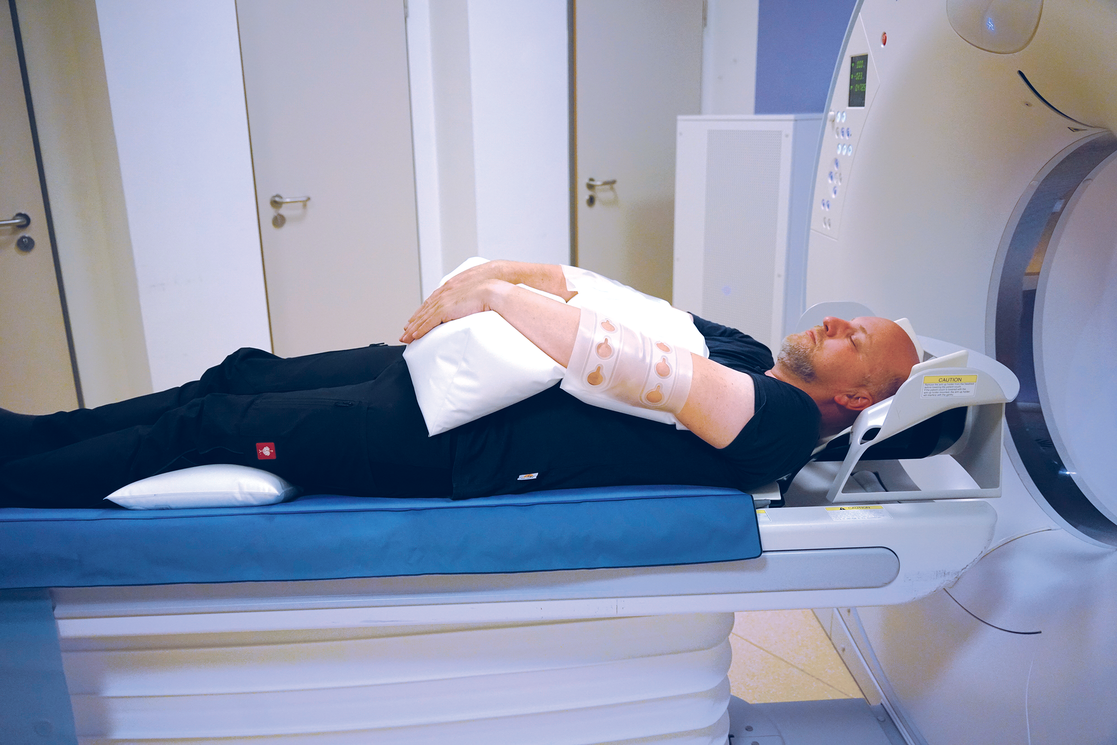
- head first supine. Epicondyles medial and lateral are aligned and straight. Minimizes radiation to patients head.
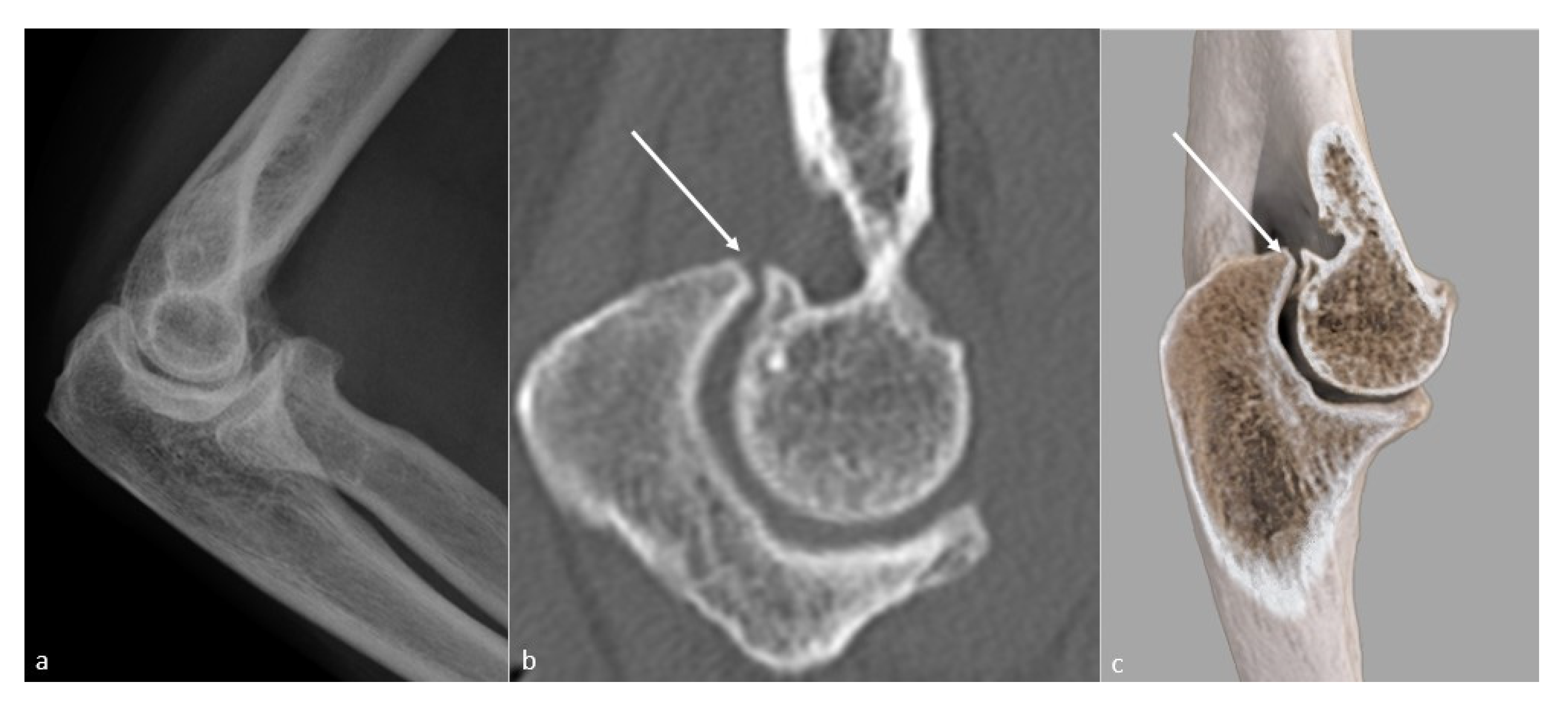
Visible in two different windowing scans bone and soft tissues and in three different planes axial coronal and sagittal. Noncontrast NA Same Same Do not repeat CT scan recon soft tissue from 1st acquisition send soft tissue kernal volume to TeraRecon Use 4mm corsag if large patient or metal in FOV. The patient is told to maintain the position while the table moves a short distance and is instructed to move his body along with the table.
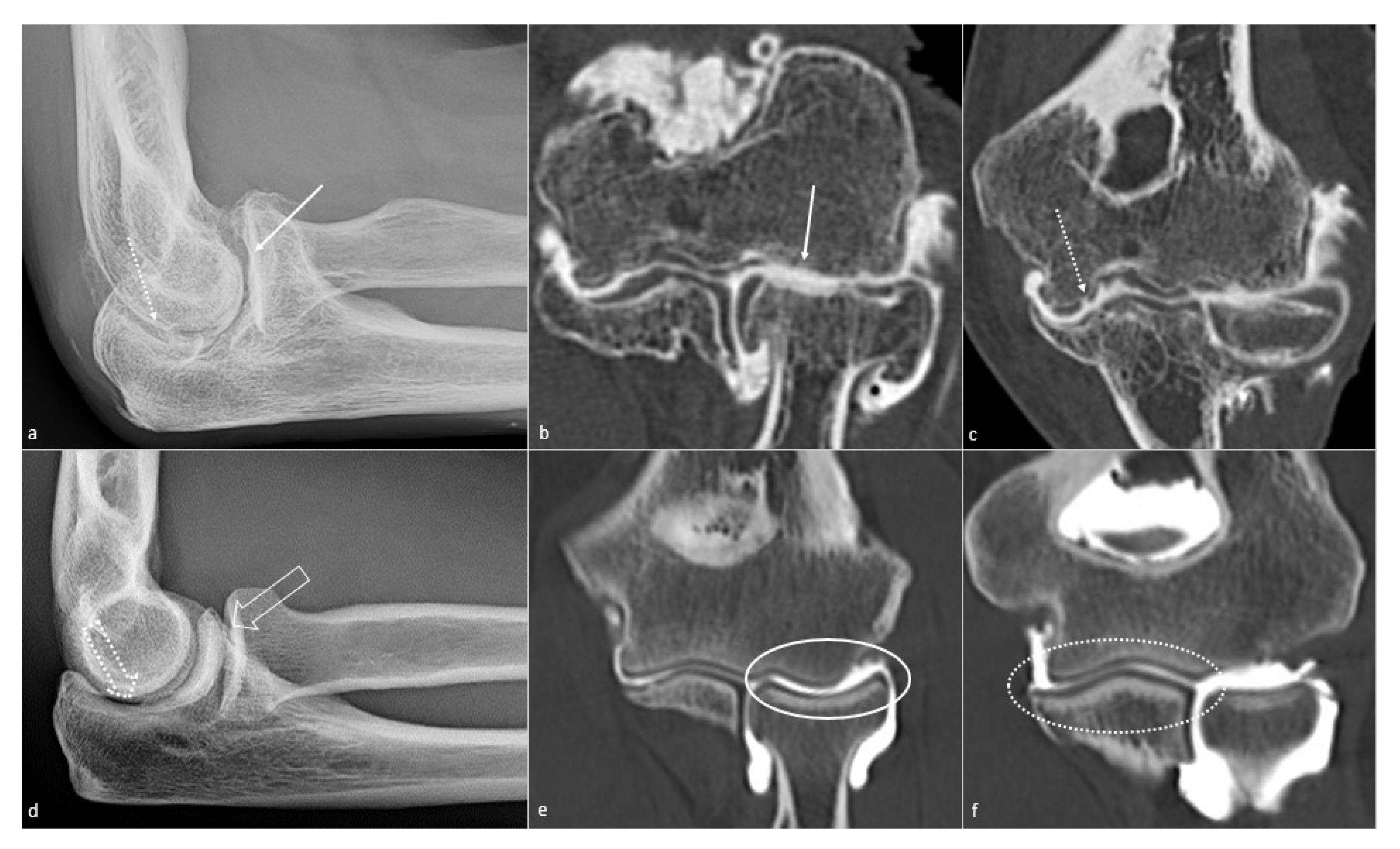
Different CT protocols have been described and CT measurement of RUI has been shown to be susceptible to changes in elbow positioning particularly pronation and. Radiology department Rijnland Hospital Leiderdorp the Netherlands.
When assessing a cranio-caudal elbow image the key indicators of good positioning are.
See illustration - Distal humeral shaft to just past radial tuberosity Series Name. Reformat in 6 planes. CT scout 1605 1739 CT. See illustration - Distal humeral shaft to just past radial tuberosity Series Name. Variation in antebrachium positioning did yield a difference in congruence. Elbow fractures in Children. The assessment of the elbow can be difficult because of the changing anatomy of the growing skeleton and the subtility of some of. The Olecranon should be superimposed over the supratochlear foramen. Coronal Imaging Plane Prescribe plane parallel to anterior humerus at condyles.
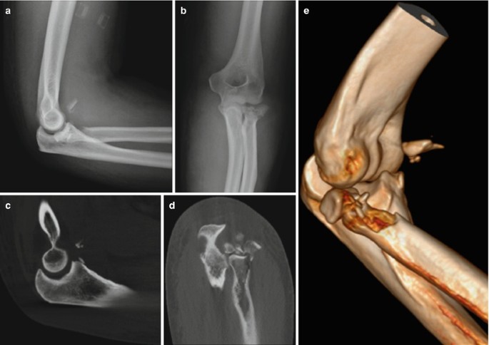
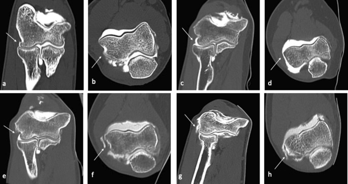
When assessing a cranio-caudal elbow image the key indicators of good positioning are. Minimizes radiation to patients head. Elbow fractures in Children. Ideally the upper arm elbow and forearm are all resting on the table. A bone algorithm of. Different CT protocols have been described and CT measurement of RUI has been shown to be susceptible to changes in elbow positioning particularly pronation and. CT imaging found that two of these patients had a radial head fracture two others had an olecranon fracture and one patient had a coronoid.



















Post a Comment for "Ct Elbow Positioning"